Cortical layers
Cortical layer markers provide a useful tool for studying the development, functional neuroanatomy and pathology of the cerebral cortex.
Atlas Antibodies offers a wide range of neuroinflammation markers. Their antibodies are affinity-purified, reproducible, and selective for their target proteins through an enhanced validation process.
Neuroinflammation broadly defines the collective reactive immune response in the brain and spinal cord in response to injury and disease.
Neuroinflammation has been implicated in acute brain trauma and hypoxic-ischemic brain damage following stroke, chronic infection and neurodegenerative diseases, such as Alzheimer’s disease, amyotrophic lateral sclerosis, Lewy body dementia, and leukoencephalopathies like multiple sclerosis.
Atlas Antibodies offers a wide range of neuroinflammation markers targeting non-neuronal cell types involved in neuroinflammation processes such as microglia, oligodendrocytes, and astrocytes.
Glial cells, comprising astrocytes, oligodendrocytes, and microglia, are the non-neuronal cell population of the central nervous system (CNS). Glial cells do not produce electrical impulses. They maintain homeostasis, form myelin, and provide support and protection for neurons. Neuroinflammation arises within the CNS through phenotypic changes of the glial cells causing the release of different cytokines and chemokines, loss of myelin and neurons, recruitment, and infiltration of peripheral blood cells, mainly T-and B-cells, into the brain parenchyma.
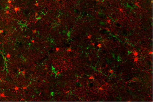
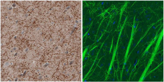
Astrocytes are the most abundant glial cell type in the CNS and the crucial regulators of innate and adaptive immune responses. Astrocytes regulate the formation, maturation, maintenance, and stability of synapses by secreting various molecules critical for synaptogenesis. Astrocytes also contribute to the induction and maintenance of the blood-brain barrier. In response to neuroinflammation, they undergo a process termed astrogliosis, which results in a spectrum of heterogenous changes that vary with etiology and severity of CNS injury. Classically, this process is characterized by upregulation of GFAP and vimentin, key astrocyte intermediate filaments, and hypertrophy of astrocyte processes. Antibodies targeting astrocytes include the Anti-GFAP, Anti-VIM, Anti-GLUL, Anti-S100B, Anti-EZR, and Anti-MT3.

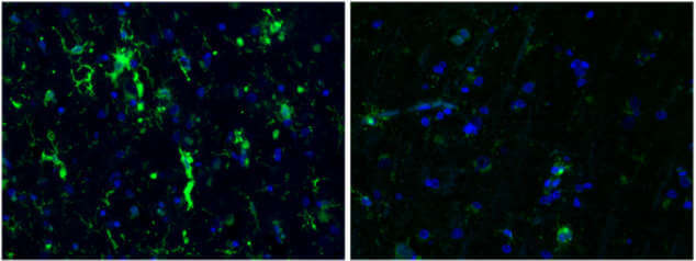
Microglia cells are critical cellular mediators of neuroinflammatory processes. As the resident macrophage cells, they act as the first and main form of active immune defense in the CNS. Activation of microglia is a hallmark of brain pathology. The chronic activation of microglia may cause neuronal damage through the release of potentially cytotoxic molecules such as proinflammatory cytokines, reactive oxygen intermediates, proteinases, and complement proteins. For example, in the pathogenesis of Alzheimer´s disease, activated microglia participates in amyloid Aβ-plaques formation and the associated pathology. Antibodies targeting microglia cells include the Anti-AIF, Anti-CD68, Anti-HLA-DRA, Anti-ITGAM (CD11b), Anti-TMEM119, AIF1/Iba1-2, Anti-P2RY12, and Anti-PTPRC.

Oligodendrocytes are highly specialized glial cells whose function is to myelinate the CNS axons (equivalent to the function performed by Schwann cells in the peripheral nervous system). Myelination of axons is fundamental for the rapid conduction of nerve impulses and contributes to axonal integrity. The devastating neurological deficits caused by demyelinating diseases, such as multiple sclerosis, clearly illustrate the importance of well-functioning oligodendrocytes. Antibodies targeting oligodendrocytes include the Anti-MBP, Anti-MOG, Anti-CNP, Anti-GRP17, and Anti-OlIg2.

Here you can find a selection of Atlas Antibodies’ PrecisA™ monoclonal antibodies targeting neuroinflammation processes.
We gladly support you by keeping you updated on our latest products and the developments around our services.