Making the ideal antibody conjugate has never been easier!
Easily label your own antibody with a selection of over 30 CF® Dye labels using Mix-n-Stain™ Antibody Labeling Kits
Published in over 200 studies, NucView® Caspase-3 Substrates offer a validated solution for real-time monitoring of apoptosis in live cells by microscopy or flow cytometry.
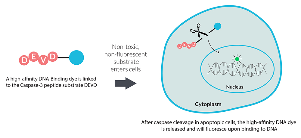
NucView® Caspase-3 Substrates are based on novel fluorogenic DNA dyes that have been coupled to the caspase-3/7 recognition sequence (DEVD). When introduced to cells, the substrate is initially non-fluorescent and is able to penetrate the plasma membrane and enter the cytoplasm. During apoptosis, caspase-3/7 cleaves the substrate and releases the high-affinity DNA dye which leads to nuclear fluorescent staining. Consequently, NucView® Caspase-3 Substrates are bifunctional, allowing detection of caspase-3/7 activity and visualization of morphological changes in the nucleus during apoptosis.

Principle of NucView® substrate technology. The substrate is non-fluorescent and does not bind DNA. After enzyme cleavage, the high-affinity DNA dye is released and fluoresces upon binding to DNA.
In contrast to other fluorogenic caspase substrates or similar assays, NucView® Caspase-3 Substrates can be used to detect caspase-3/7 activity in live cells without inhibiting apoptosis progression. Staining is also compatible with subsequent fixation and permeabilization for downstream immunostaining.
Explore assay kits that combine the advantage of NucView® real-time apoptosis analysis with other probes for monitoring necrosis, membrane potential, or other apoptosis markers.
Learn more about NucView® Caspase-3 Substrates ↗
Download the E-Book “Visualizing Apoptosis as it Happens” to understand how caspase 3/7 activity plays a role in the mechanism underlying programmed cell death.

We gladly support you by keeping you updated on our latest products and the developments around our services.