Neuroinflammation monoclonal antibodies
Atlas Antibodies offers a wide range of neuroinflammation markers. Their antibodies are affinity-purified, reproducible, and selective for their target proteins through an enhanced validation process.
Cortical layer markers provide a useful tool for studying the development, functional neuroanatomy and pathology of the cerebral cortex.
Cerebral cortex is the part of the human brain that undergoes the most profound evolutional changes and serves as a substrate for the higher cognitive functions. During embryonal development, six distinct layers are generated from the progenitors of the neocortical germinal zone. In the adult brain the different cortical layers are defined based on morphologically and functionally divergent neurons.

The six cortical layers are generated during embryonal development in a strictly regulated manner:
Following formation of the cortical plate, the deeper layers neurons are generated (including layers V and VI) and thereafter the upper layers neurons (layers II-IV). Glial cells, including astrocytes and oli-godendrocytes, are then generated at the latest stages of cortical development.
The neocortical neurogenesis is depend-ent on several transcriptional factors. For instance, LHX2 and PAX6 together play a crucial role in the specification of neo-cortical progenitors which give rise to the projection neurons. MEF2C is another example of transcription factor essential for normal neural development and spatial distribution in the neocortex. Figure 1 shows expression profiles of LHX2, PAX6 and MEF2C in the developing mouse brain.

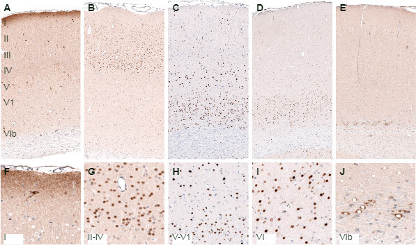
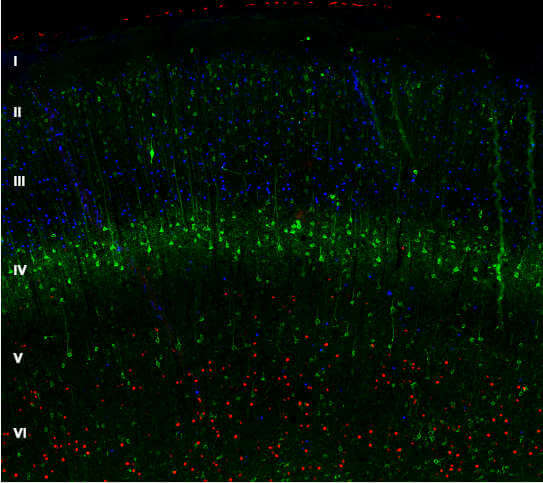
In the mature neocortex, neurons of different layers display anatomical and functional diversity, including cell morphology, physiological properties and anatomical connections. Neurons of layers II/III, along with a subset of neurons of layer V, contribute mostly to intracortical connections, including the callosal projections to the contralateral cerebral hemisphere. Layer V corticofugal neurons target the midbrain, hindbrain and spinal cord, while layer VI neurons project mainly to the thalamus. Protein expression profiles differ in neurons of various layers. For example, upper layers neurons can be identified by expression of CUX1 and POU3F2 (BRN2), the neurons of layer V – by expression of BCL11B (CTIP2) and neurons of layer VI – by FOXP2 expression. Laminar distribution of these and some other markers in rat neocortex is shown in Figure 2 and Figure 3.

| Cortical Layer | Product Name |
|---|---|
| Layer 1 | Anti-RELN (AMAb91365) |
| Layer 1 | Anti-RELN (HPA046512) |
| Layer 2/3 | Anti-RASGRF2 (HPA018679) |
| Layer 2/3 | Anti-CALB1 (HPA023099) |
| Layer 2/3-4 | Anti-CUX1 (AMAb91352) |
| Layer 2/3-4 | Anti-CUX1 (AMAb91353) |
| Layer 2/3-4 | Anti-CUX1 (HPA003277) |
| Layers 2/3, 4 and 5b | Anti-POU3F2 (BRN2) (HPA056261) |
| Layers 2/3, 4 and 5b | Anti-POU3F2 (BRN2) (AMAb91406) |
| Layers 2/3, 4 and 5b | Anti-POU3F2 (BRN2) (AMAb91407) |
| Layer 2-4 (mainly 4) | Anti-NECAB1 (AMAb90798) |
| Layer 2-4 (mainly 4) | Anti-NECAB1 (AMAb90800) |
| Layer 2-4 (mainly 4) | Anti-NECAB1 (AMAb90801) |
| Layer 2-4 (mainly 4) | Anti-NECAB1 (HPA023629) |
| Layer 2-4 (mainly 4) | Anti-NECAB1 (HPA031262) |
| Layer 5 | Anti-PCP4 (HPA005792) |
| Layer 5 | CNTN6 (HPA016645) |
| Layers 5-6 | Anti-BCL11B(CTIP2) (HPA049117) |
| Layer 6 | Anti-FOXP2 (AMAb91361) |
| Layer 6 | Anti-FOXP2 (AMAb91362) |
| Layer 6 | Anti-FOXP2 (HPA000382) |
| Layer 6 | Anti-TLE4 (HPA065357) |
| Layer 6b | Anti-CTGF (AMAb91366) |
| Layer 6b | Anti-CTGF (HPA031075) |
We gladly support you by keeping you updated on our latest products and the developments around our services.