Biochemical and cell-based assays to interrogate ADP-ribosylation
BPS Bioscience supports scientific discovery with innovative research solutions. Our off-the-shelf assay kits facilitate research inquiries as well as drug discovery & development efforts.
Biochemical and cell-based assays to interrogate ADP-ribosylation
The reversible addition of ADP-ribose units to proteins is a vital post-translational modification involved in cellular processes such as DNA repair, chromatin remodeling, and control of gene expression. It happens in proteins linked to the DNA Damage Response (DDR). This addition comes in two flavors: mono-ADP-ribosylation (a single ADP-ribose unit is added, also termed MARylation) and poly-ADP-ribosylation (multiple units are added in linear or branched forms), also known as PARylation. These reactions are orchestrated by a family of seventeen enzymes known as poly (ADP-ribose) polymerases (PARPs), with PARP1 to PARP5 catalyzing PARylation, and the other PARPs performing MARylation. PARP1/2 inhibitors have been game-changing in cancer therapy, demonstrating the potential efficacy of synthetic lethal drugs targeting complementary pathways within the DNA damage response network. Indeed, there are now four FDA-approved drugs in clinical use in the US, with many more in the pipeline.
On the other hand, removal of ADP-ribose from proteins is performed by poly (ADP-ribose) glycohydrolase (PARG) and ADP-ribosyl-acceptor hydrolase 3 (ARH3), which participate in cancer progression and are also considered promising therapeutic targets.
Choose your assay kit depending on your objective
Whether you aim at measuring levels of PARylation from intact cells, PARG/ARH3 or PARP enzymatic activity for drug profiling, or at evaluating a compound ability to trap PARP onto DNA, there is an assay for you. Our assay kits contain all the reagents required to perform the experiment, including ECL reagents where necessary. To compare the effect of a compound on the entire PARP family, inquire about custom services.
Evaluate cellular PARylation levels
Cell-based assays are an essential part of the drug development process, as they allow researchers to evaluate the biological activity of drug candidates and gain a better understanding of their mechanism of action. Cell assays are necessary to evaluate the compounds’ membrane permeability issues and interferences within a complex system, and overall provide a more accurate representation of the complex interactions between compounds and living cells, as compared to biochemical assays. The LysA™ Universal PARylation Assay Kit evaluates PARylation levels in cellular extracts and allows determination of compound IC50, comparison in the IC50 of various compounds, the effect of a compound in cells with specific DDR defects, and more.
Example applications
Measure total PAR levels after cell stress induced by chemotherapy drug (indicates, for example, if PARP was activated and to what extent).
Screen or determine the IC50 of inhibitors of the PARP family (PAR writers).
Screen or determine the IC50 of inhibitors of the PARG family (PAR erasers).
Validate a new cellular model for the study of PAR homeostasis.
Evaluate the possible synergistic effect of treatment combinations.
Quantify PAR levels in biological samples using a defined PAR standard.
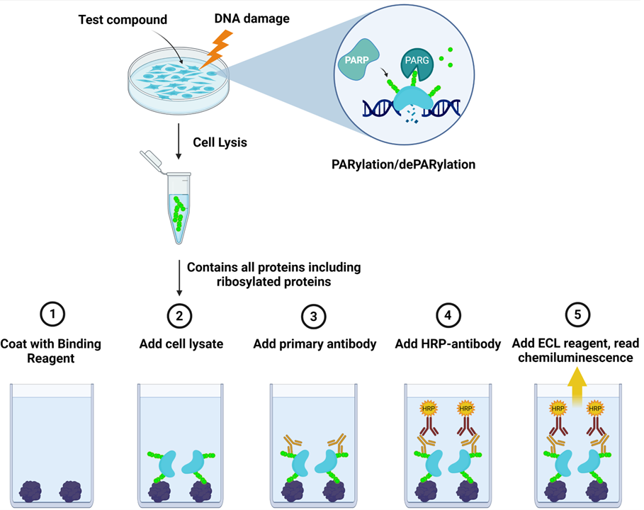
Assay principle: The LysA™ Universal PARylation Assay Kit (#82123) is a sandwich ELISA-based assay designed to analyze the level of total PARylation present in cellular extracts. The kit includes a PAR standard for absolute quantification. The assay detects differences in protein PARylation levels resulting, for example, from inducing the DNA damage response or from exposure to PARP inhibitors or to PARG inhibitors. A 96-well plate is coated with a binding reagent that recognizes PARylated chains. Lysates from cells are added to the coated wells, and PARylated proteins present in the cell lysates are captured by the Binding Reagent. This is followed by an incubation with an anti-PAR detection antibody, then a secondary HRP-conjugated antibody. Addition of a chemiluminescent HRP substrate provides a luminescence signal that directly correlates with the level of cellular PARylation. Of note, the assay does not detect mono-ADP-ribosylation (MARylation).
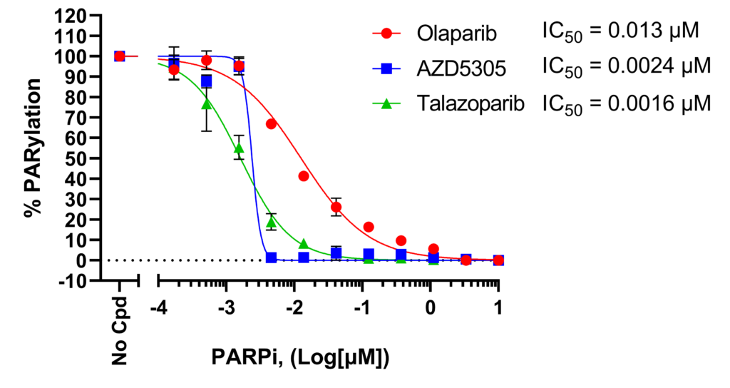
Effect of PARP inhibitors on H2O2-induced protein PARylation in HEK293 cells.
Cells were treated with increasing concentrations of PARP inhibitors before inducing DNA damage with H2O2 and cell lysates were analyzed using LysA™ Universal PARylation Assay Kit. Results were normalized as percent of total PARylation in which maximum PARylation was set to 100%. For each condition, the IC50 was calculated and plotted using GraphPad Prism software.
Measure PARP enzymatic activity in vitro
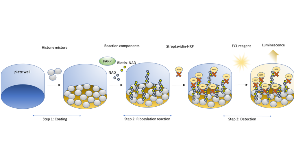
These chemiluminescent ELISA use purified PARP proteins to measure the effect of test compounds on the enzymatic activity of the proteins, i.e., the PARylation or MARylation of a histone substrate. Briefly, histone proteins are coated on a 96-well plate. A biotinylated NAD+ mix is incubated with the purified PARP enzyme. Depending on the enzyme, an activated DNA template may be added. After washing, streptavidin-HRP is added, the plate is washed again, followed by addition of an ECL substrate to produce a chemiluminescence signal that is proportional to PARP activity.
It has been shown that some PARP1/2 inhibitors act by preventing PARP auto-PARylation, causing failure to detach from the DNA and effectively trapping PARP, preventing repair, and leading to cell death. Thus, DNA trapping is a highly desirable property of PARP inhibitors. While other assays measure PARP enzymatic activity and quantify the PARylation of target proteins, such as histones, the PARPtrap™ assays measure a compound’s ability to keep PARP1 or PARP2 onto a DNA probe.
This homogeneous, simple assay can be incorporated into high-throughput drug discovery screens or rounds of optimization for molecules that enhance trapping. PARPtrap™ assays allows researchers to efficiently screen their libraries for the most effective inhibitors.
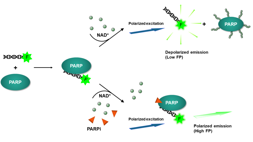
The assay is based on principles of fluorescence polarization and uses fluorescently labeled DNA probes, which, when excited by polarized light, emit fluorescence with a degree of polarization proportional to the rate of molecular rotation. Free DNA probes are very small and rotate fast, therefore they have low fluorescence polarization (FP). When in complex with PARP, molecular rotation is slow due to the large size of the protein, and fluorescence polarization is high. Upon addition of NAD+, the newly PARylated enzymes detach from the probe, reducing FP levels (see Figure 3).
In practice, the user incubates purified PARP1 or PARP2 with a specific fluorescent DNA probe, then adds NAD+ to initiate PARylation. Once PARylated, PARP detaches from the probe due to the high negative charge of the PAR chains. This is the experimental condition in which FP is the lowest since most of the DNA probe will be free. Pre-incubation of PARP with an inhibitor that blocks PARylation, but not the binding to the DNA, will keep the enzyme trapped to the DNA probe, a condition in which the probe remains engaged in a complex with PARP, resulting in high FP. Thus, the user compares a no-inhibitor control with low FP and a test inhibitor condition with high FP, which means that an increase in the FP signal indicates trapping by the inhibitor.
The PARPtrap™ assays exist in 96-well and 384-well formats for PARP1 (#80584), PARP2 (#78296), and as combo kit (#78317).
Evaluate PARG or ARH3 enzymatic activity
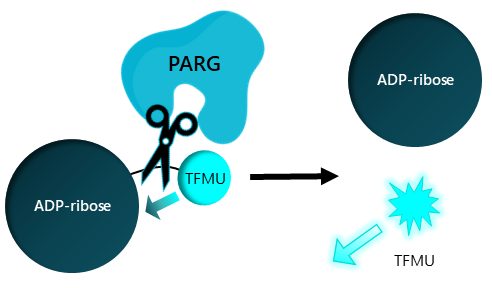
The PARG and ARH3 Fluorogenic Assay Kits are homogeneous 96-well assays designed to measure the hydrolase activity of these enzymes for screening and profiling applications, using a simple and straightforward fluorogenic assay. In these experiments, the enzyme is incubated with a fluorogenic ADP-ribose substrate in which the fluorophore is quenched by the presence of the ribose. PARG or ARH3-mediated hydrolysis of the substrate between the ribose and the fluorochrome releases fluorescence that can be detected at λ=502 nm (excitation at λ=385 nm). Fluorescence intensity is directly proportional to the level of hydrolase activity.

 Find products for your research