Illuminate your Research with Lentiviruses for Cell Biology
Simplify your genetic engineering of hard-to-transfect cells with BPS Bioscience’s lentiviral tools for imaging, cell structure visualization, and for transduction controls.
Lentiviral transduction is a powerful tool for introducing genes into cells and is used extensively for cellular engineering. It has recently made its way to the clinic with ex-vivo cell therapy applications.
How Lentiviruses Work
Lentiviruses (LVs) are RNA-based enveloped viruses that enter cells by binding to a particular cell surface protein, followed by membrane fusion. This process allows the viral RNA containing its genetic information to enter the cytoplasm. The single-stranded viral RNA is converted into DNA by reverse transcription. The viral DNA then enters the nucleus and integrates into the host cell genome with the help of a viral integrase. This mechanism is particularly useful for establishing stable modified cell lines.
HIV-Derived Lentiviruses and Modifications
The most commonly used LV is based on the RNA virus HIV (human immunodeficiency virus), which infects human lymphocytes through the specific interaction of its envelope protein gp120 with the T cell CD4 receptor. For biotechnology applications, the envelope protein gp120 has been replaced by VSV-G (Vesicular stomatitis virus G protein). VSV-G binds to the human LDL receptor (low-density lipoprotein receptor), which is expressed on most cells, making these virus particles especially versatile for targeting a wide range of cell types.
Safety and Production of Lentiviruses
Contemporary methods of LV production ensure they are very safe. A biosafety level 2 facility is sufficient for using BPS Bioscience’s lentiviruses. These replication-incompetent viral particles, produced from packaging cells, contain minimal genetic information, including the gene of interest but excluding the viral packaging genes. As a result, cells infected with these viruses cannot produce new viral particles.
BPS Bioscience Lentivirus Products
All BPS Bioscience LV products are supplied as ready-to-use viral particles that meet strict specifications for titer and size. Exact titers are provided with each lot, ensuring consistency and reliability for research and clinical applications.
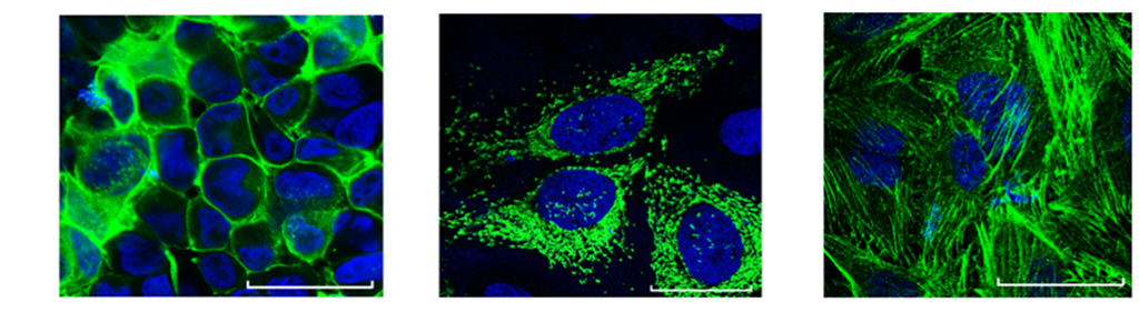
LVs for the Visualization of Subcellular Structures
BPS Bioscience new collection of lentiviruses targets subcellular structures with fluorescent proteins for direct visualization. Transduced cells express a fluorescent protein fused to a targeting sequence that directs it to a specific subcellular structure, such as the nucleus, plasma membrane, or mitochondria. These compartment-specific fluorescent proteins enable scientists to visualize the subcellular architecture with exceptional clarity using fluorescent microscopy, and facilitate live-cell imaging in real-time. By combining dual-labeling strategies, researchers can uncover protein co-localization and gain deeper insights into cellular processes. In addition, these lentiviruses support the generation of stable cell lines, ensuring consistent and reproducible expression of the fluorescently labeled proteins for long-term studies.
Expression of GFP (green fluorescent protein) at the plasma membrane (left), mitochondria (center), and β-actin (right).
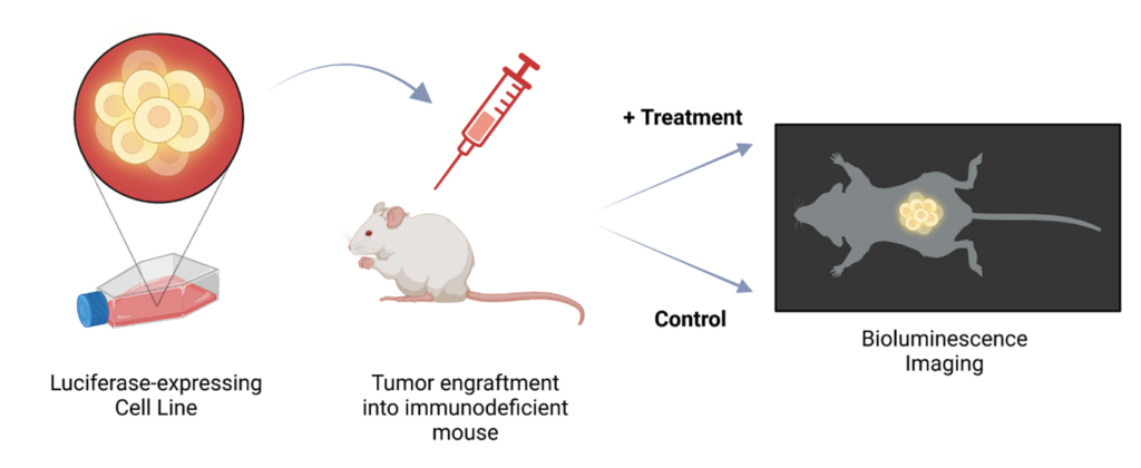
These LVs are powerful tools for expressing luminescent proteins (e.g., firefly luciferase, Renilla luciferase) or fluorescent proteins (e.g., RFP, YFP, eGFP) in cells, which then enable researchers to visualize the cells using fluorescence microscopy, analyze them with flow cytometry, or sort them via FACS (Fluorescence activated cell sorting). In vivo applications further expand their utility: fluorescently labeled cells can be observed using intra-vital microscopy, while luciferase-expressing cells are ideal for bioluminescence imaging. Thus, these reporter proteins enable dynamic tracking and functional analysis in both in vitro and in vivo systems.
Illustration of bioluminescence imaging of a mouse model generated by injection of luciferase reporter tumor cells followed by injection of In Vivo-Luc™ Imaging Solution (#78803).
Products
Firefly Luciferase Lentivirus (EF1A Promoter/Geneticin, Hygromycin, Puromycin, or Blasticidin) (#78740)
Firefly Luciferase Lentivirus (G418, Hygromycin and Puromycin) (# 79692)
A negative control LV is an essential tool in studies involving gene expression and cell line generation. Unlike experimental LVs designed to express a specific protein under a promoter, the negative control LV does not encode or express any specific protein of interest. Instead, it typically carries a selection marker—such as a fluorescent protein or antibiotic resistance gene—that allows researchers to establish a stable cell line for comparison.
The primary purpose of a negative control is to serve as a baseline or reference. When generating a cell line using lentiviral transduction, variations in cellular behavior or responses could result from the transduction process itself, rather than from the expression of the target protein. The negative control ensures that any observed differences in experimental outcomes can be attributed to the protein of interest rather than nonspecific effects of the lentiviral vector, promoter activity, or the selection process.
BPS Bioscience provides a collection of control LVs containing antibiotic resistance genes commonly used in cellular engineering, such as geneticin, puromycin, or hygromycin resistance genes.
Products
Expression Negative Control Lentivirus (Inducible TET On™) (#82290)
Expression Negative Control Lentivirus (EF1A Promoter/Hygromycin, Puromycin, or G418) (# 82212)
Expression Negative Control Lentivirus (G418 or Hygromycin or Puromycin) (#79902)
Negative Control eGFP Reporter Lentivirus (#79927)

 Find products for your research